


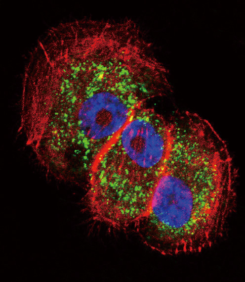
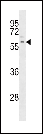

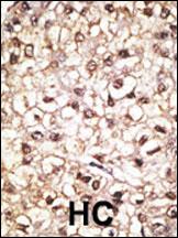
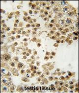
Application 
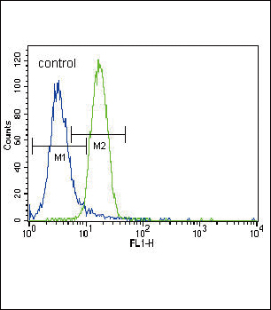
| WB, IHC-P, IF, FC, E |
|---|---|
| Primary Accession | O60260 |
| Other Accession | NP_004553 |
| Reactivity | Human, Mouse |
| Host | Rabbit |
| Clonality | Polyclonal |
| Isotype | Rabbit IgG |
| Calculated MW | 51641 Da |
| Antigen Region | 387-417 aa |
| Gene ID | 5071 |
|---|---|
| Other Names | E3 ubiquitin-protein ligase parkin, 632-, Parkinson juvenile disease protein 2, Parkinson disease protein 2, PARK2, PRKN |
| Target/Specificity | This Parkin antibody is generated from rabbits immunized with a KLH conjugated synthetic peptide between 387-417 amino acids from the C-terminal region of human Parkin. |
| Dilution | IF~~1:10~50 WB~~1:1000 IHC-P~~1:50~100 FC~~1:10~50 |
| Format | Purified polyclonal antibody supplied in PBS with 0.09% (W/V) sodium azide. This antibody is purified through a protein A column, followed by peptide affinity purification. |
| Storage | Maintain refrigerated at 2-8°C for up to 2 weeks. For long term storage store at -20°C in small aliquots to prevent freeze-thaw cycles. |
| Precautions | Parkin Antibody (C-term) is for research use only and not for use in diagnostic or therapeutic procedures. |
| Name | PRKN (HGNC:8607) |
|---|---|
| Synonyms | PARK2 |
| Function | Functions within a multiprotein E3 ubiquitin ligase complex, catalyzing the covalent attachment of ubiquitin moieties onto substrate proteins (PubMed:10888878, PubMed:10973942, PubMed:11431533, PubMed:12150907, PubMed:12628165, PubMed:15105460, PubMed:16135753, PubMed:21376232, PubMed:21532592, PubMed:22396657, PubMed:23620051, PubMed:23754282, PubMed:24660806, PubMed:24751536, PubMed:29311685, PubMed:32047033). Substrates include SYT11 and VDAC1 (PubMed:29311685, PubMed:32047033). Other substrates are BCL2, CCNE1, GPR37, RHOT1/MIRO1, MFN1, MFN2, STUB1, SNCAIP, SEPTIN5, TOMM20, USP30, ZNF746, MIRO1 and AIMP2 (PubMed:10888878, PubMed:10973942, PubMed:11431533, PubMed:12150907, PubMed:12628165, PubMed:15105460, PubMed:16135753, PubMed:21376232, PubMed:21532592, PubMed:22396657, PubMed:23620051, PubMed:23754282, PubMed:24660806, PubMed:24751536). Mediates monoubiquitination as well as 'Lys-6', 'Lys-11', 'Lys-48'-linked and 'Lys-63'-linked polyubiquitination of substrates depending on the context (PubMed:19229105, PubMed:20889974, PubMed:25474007, PubMed:25621951, PubMed:32047033). Participates in the removal and/or detoxification of abnormally folded or damaged protein by mediating 'Lys-63'-linked polyubiquitination of misfolded proteins such as PARK7: 'Lys-63'-linked polyubiquitinated misfolded proteins are then recognized by HDAC6, leading to their recruitment to aggresomes, followed by degradation (PubMed:17846173, PubMed:19229105). Mediates 'Lys-63'-linked polyubiquitination of a 22 kDa O-linked glycosylated isoform of SNCAIP, possibly playing a role in Lewy-body formation (PubMed:11431533, PubMed:11590439, PubMed:15105460, PubMed:15728840, PubMed:19229105). Mediates monoubiquitination of BCL2, thereby acting as a positive regulator of autophagy (PubMed:20889974). Protects against mitochondrial dysfunction during cellular stress, by acting downstream of PINK1 to coordinate mitochondrial quality control mechanisms that remove and replace dysfunctional mitochondrial components (PubMed:11439185, PubMed:18957282, PubMed:19029340, PubMed:19966284, PubMed:21376232, PubMed:22082830, PubMed:22396657, PubMed:23620051, PubMed:23933751, PubMed:24660806, PubMed:24784582, PubMed:24896179, PubMed:25474007, PubMed:25527291, PubMed:32047033). Depending on the severity of mitochondrial damage and/or dysfunction, activity ranges from preventing apoptosis and stimulating mitochondrial biogenesis to regulating mitochondrial dynamics and eliminating severely damaged mitochondria via mitophagy (PubMed:11439185, PubMed:19029340, PubMed:19801972, PubMed:19966284, PubMed:21376232, PubMed:22082830, PubMed:22396657, PubMed:23620051, PubMed:23685073, PubMed:23933751, PubMed:24896179, PubMed:25527291, PubMed:32047033, PubMed:33499712). Activation and recruitment onto the outer membrane of damaged/dysfunctional mitochondria (OMM) requires PINK1-mediated phosphorylation of both PRKN and ubiquitin (PubMed:24660806, PubMed:24784582, PubMed:25474007, PubMed:25527291). After mitochondrial damage, functions with PINK1 to mediate the decision between mitophagy or preventing apoptosis by inducing either the poly- or monoubiquitination of VDAC1, respectively; polyubiquitination of VDAC1 promotes mitophagy, while monoubiquitination of VDAC1 decreases mitochondrial calcium influx which ultimately inhibits apoptosis (PubMed:27534820, PubMed:32047033). When cellular stress results in irreversible mitochondrial damage, promotes the autophagic degradation of dysfunctional depolarized mitochondria (mitophagy) by promoting the ubiquitination of mitochondrial proteins such as TOMM20, RHOT1/MIRO1, MFN1 and USP30 (PubMed:19029340, PubMed:19966284, PubMed:21753002, PubMed:22396657, PubMed:23620051, PubMed:23685073, PubMed:23933751, PubMed:24896179, PubMed:25527291). Preferentially assembles 'Lys-6'-, 'Lys-11'- and 'Lys-63'-linked polyubiquitin chains, leading to mitophagy (PubMed:25621951, PubMed:32047033). The PINK1-PRKN pathway also promotes fission of damaged mitochondria by PINK1-mediated phosphorylation which promotes the PRKN-dependent degradation of mitochondrial proteins involved in fission such as MFN2 (PubMed:23620051). This prevents the refusion of unhealthy mitochondria with the mitochondrial network or initiates mitochondrial fragmentation facilitating their later engulfment by autophagosomes (PubMed:23620051). Regulates motility of damaged mitochondria via the ubiquitination and subsequent degradation of MIRO1 and MIRO2; in motor neurons, this likely inhibits mitochondrial intracellular anterograde transport along the axons which probably increases the chance of the mitochondria undergoing mitophagy in the soma (PubMed:22396657). Involved in mitochondrial biogenesis via the 'Lys-48'-linked polyubiquitination of transcriptional repressor ZNF746/PARIS which leads to its subsequent proteasomal degradation and allows activation of the transcription factor PPARGC1A (PubMed:21376232). Limits the production of reactive oxygen species (ROS) (PubMed:18541373). Regulates cyclin-E during neuronal apoptosis (PubMed:12628165). In collaboration with CHPF isoform 2, may enhance cell viability and protect cells from oxidative stress (PubMed:22082830). Independently of its ubiquitin ligase activity, protects from apoptosis by the transcriptional repression of p53/TP53 (PubMed:19801972). May protect neurons against alpha synuclein toxicity, proteasomal dysfunction, GPR37 accumulation, and kainate-induced excitotoxicity (PubMed:11439185). May play a role in controlling neurotransmitter trafficking at the presynaptic terminal and in calcium-dependent exocytosis. May represent a tumor suppressor gene (PubMed:12719539). |
| Cellular Location | Cytoplasm, cytosol. Nucleus. Endoplasmic reticulum. Mitochondrion. Mitochondrion outer membrane {ECO:0000250|UniProtKB:Q9WVS6}. Cell projection, neuron projection. Postsynaptic density {ECO:0000250|UniProtKB:Q9WVS6}. Presynapse {ECO:0000250|UniProtKB:Q9WVS6}. Note=Mainly localizes in the cytosol (PubMed:19029340, PubMed:19229105). Co-localizes with SYT11 in neutrites (PubMed:12925569). Co-localizes with SNCAIP in brainstem Lewy bodies (PubMed:10319893, PubMed:11431533). Translocates to dysfunctional mitochondria that have lost the mitochondrial membrane potential; recruitment to mitochondria is PINK1-dependent (PubMed:18957282, PubMed:19966284, PubMed:23620051, PubMed:24898855) Mitochondrial localization also gradually increases with cellular growth (PubMed:22082830). |
| Tissue Location | Highly expressed in the brain including the substantia nigra (PubMed:19501131, PubMed:9560156). Expressed in heart, testis and skeletal muscle (PubMed:9560156). Expression is down- regulated or absent in tumor biopsies, and absent in the brain of PARK2 patients (PubMed:12719539, PubMed:14614460). Overexpression protects dopamine neurons from kainate-mediated apoptosis (PubMed:12628165) Found in serum (at protein level) (PubMed:19501131) |