Goat anti-Rabbit IgG (H&L), ALP conjugated
Cat# AS09607
Size : 1mg
Brand : Agrisera
1
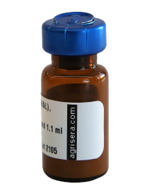
AS09 607 | Clonality: Polyclonal Host: Goat Reactivity: Rabbit IgG (H&L)
- Product Info
-
Immunogen: Purified Rabbit IgG, whole molecule Host: Goat Clonality: Polyclonal Purity: Immunogen affinity purified goat IgG. Format: Liquid, clear, colorless. Quantity: 1 mg Storage: Non-diluted antibody is stable for 4 years at 2-8°C. For storage at -20°C dilute antibody solution with an equal volume of glycerol to obtain final glycerol concentration of 50 % to prevent loss of enzymatic activity. Such solution will not freeze in -20°C. If you are using a 1:5000 dilution prior to diluting with glycerol, then you would need to use a 1:2500 dilution after adding glycerol. Prepare working dilution prior to use and then discard, Be sure to mix well but without foaming. Tested applications: ELISA (ELISA), Immunohistochemistry (IHC), Western blot (WB) Recommended dilution: 1 : 500-1: 8 000 (ELISA), 1 : 500 -1 : 2000 (IHC), 1 : 500-1: 8 000 (WB) - Reactivity
-
Confirmed reactivity: Rabbit IgG heavy and light chains (H&L) Not reactive in: No confirmed exceptions from predicted reactivity are currently known - Application Examples
-
Application example
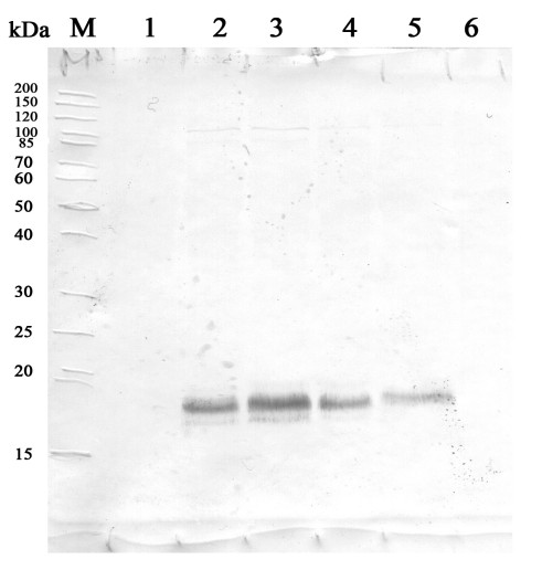
24 µg of Triticum aestivum L. whole leaf extract (1), 23 µg of Triticum aestivum L. whole leaf extract 37°С,3h (2), 22 µg of Triticum aestivum L. whole leaf extract, 37°С, 24h (3), 20 µg of Triticum aestivum L. whole leaf extract, 37°С,24h+50°C/1h (4), 17 µg of Triticum aestivum L. whole leaf extract, 37°С,24h+50°C, 3h (5), 23 µg of Triticum aestivum L. whole leaf extract, Control+50°C, 1h (6).
700 µg of total protein from spring wheat Triticum aestivum L. green leaves extracted with write exact buffer components 100 mM Tris HCl (pH=7.4), 1 mM beta-mercaptoethanol, 1 mM PMSF and denatured with 65.2 mM Tris HCl (pH=6.8), 1mM EDTA, 1% SDS, 20% glycerol, 5% beta-mercaptoethanol at 97°C for 5 min and 20 μg of total protein were separated on 12.5 % SDS-PAGE and blotted 2h on nitrocellulose membrane (GE Healthcare) using tank transfer. Blots were blocked with a skimmed milk 3 % in TBS for 1h at room temperature (RT) with agitation. Blot was incubated in the primary antibody at a dilution of 1: 1 000 for 1h at RT with agitation in TBS. The antibody solution was decanted and the blot washed 2 times for 10 min in TBS-T at RT with agitation. Blot was incubated in secondary antibody (anti-rabbit IgG, ALP conjugated, AS09 607, from Agrisera) diluted to 1:1000 in a skimmed milk 3 % in TBS for 1h at RT with agitation. The blot was washed 3 times for 5 min in TBS-T at RT with agitation and developed WB. The proteins were detected with 5-bromo-4-chloro-3-indolyl phosphate (Thermo Scientific) and Nitrotetrazolium Blue (Thermo Scientific). Exposure time was 5.20 minutes.
Courtesy of Dr. Olga Borovik, Laboratory of Physiological Genetics Siberian Institute of Plant Physiology and Biochemistry SB RAS RussiaApplication examples: 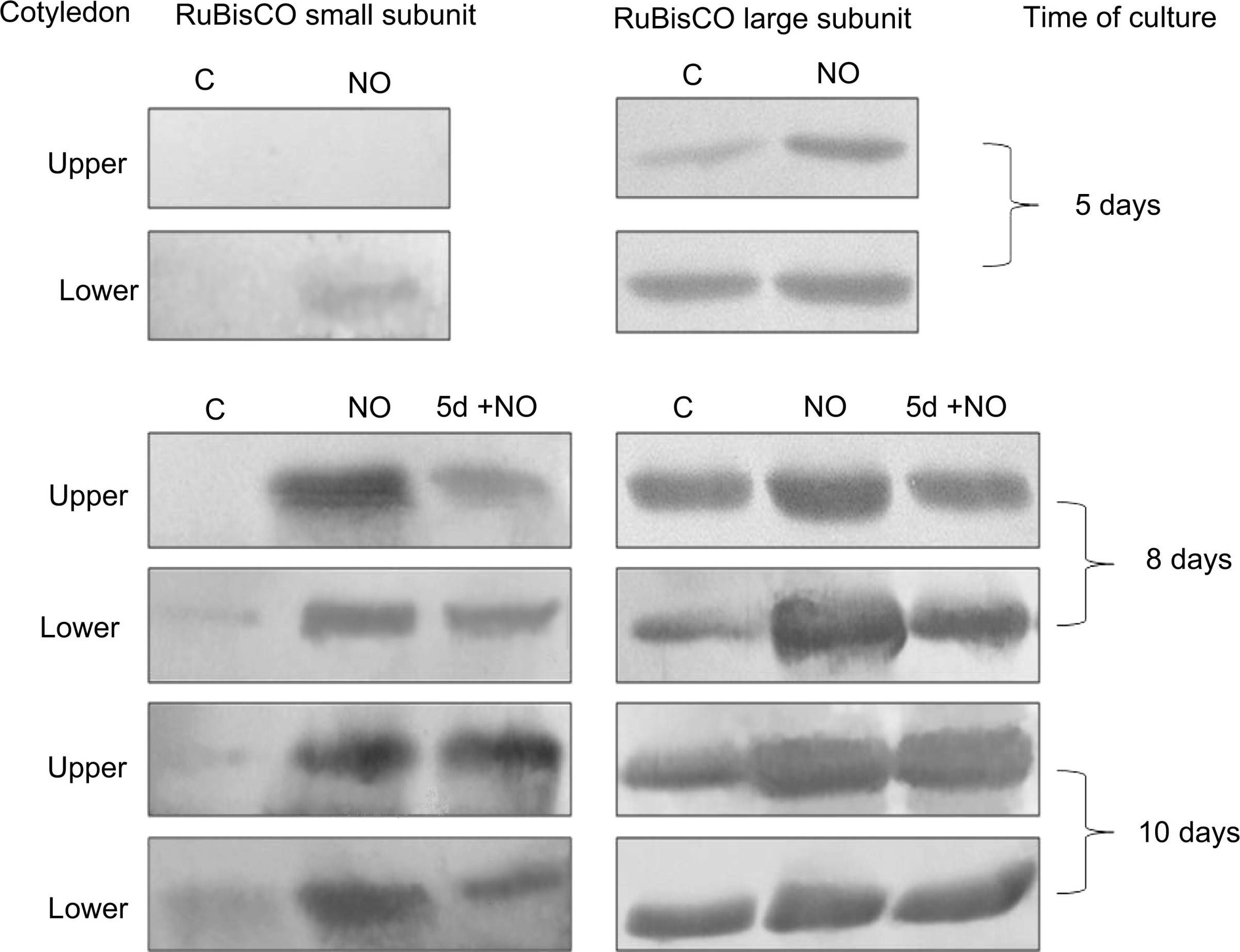
Reactant: Malus domestica (Apple)
Application: Western Blotting
Pudmed ID: 26186967
Journal: Planta
Figure Number: 6A
Published Date: 2015-11-01
First Author: Gniazdowska, A.
Impact Factor: 4.561
Open PublicationWestern blot analysis of RuBisCO large and small subunits in upper and lower cotyledons of 5-, 8-, 10-day-old control seedlings (C), cotyledons isolated from seedlings developed from embryos shortly treated with NO (NO) after 5, 8 and 10 days of culture, and cotyledons isolated from control seedlings treated with NO after 5 days of culture (5 + NO) after 8 and 10 days of experiment. Typical blots are shown
- Additional Information
-
Additional information: Concentration: 1.5mg/ml.
Antibody has been affinity purified on solid phase rabbit IgG (H&L).
AP conjugate is supplied in 30 mM Triethanolamine, pH 7.2, 5 mM Magnesium Chloride, 0.1 mM Zinc Chloride, 1 % (w/v) BSA, Protease/IgG free. 0.05 % (w/v) of sodium azide is added as preservative
Additional information (application): Based upon IEP, this antibody binds to:
• Heavy chains on rabbit IgG
• Light chains on all rabbit immunoglobulins
No reactivity is observed to non-immunoglobulin rabbit serum proteins based in immunoelectrophoresis. - Background
-
Background: Goat anti-rabbit IgG (H&L) is a secondary antibody conjugated to AP or ALP (Alkaline phosphatase) which binds to all rabbit immunoglobulins in immunological assays.
- Product Citations
-
Selected references: Rodrigues et al (2022) Exploring the Applicability of Calorespirometry to Assess Seed Metabolic Stability Upon Temperature Stress Conditions-Pisum sativum L. Used as a Case Study. Front Plant Sci. 2022 Apr 27;13:827117. doi: 10.3389/fpls.2022.827117. PMID: 35574105; PMCID: PMC9094064.
Hura et al. (2022) Physiological and molecular features predispose native and invasive populations of sweet briar (Rosa rubiginosa L.) to colonization and restoration of drought degraded environments, Perspectives in Plant Ecology, Evolution and Systematics, Volume 56, 2022, 125690, ISSN 1433-8319, https://doi.org/10.1016/j.ppees.2022.125690.
Loudya et al. (2021) Cellular and transcriptomic analyses reveal two-staged chloroplast biogenesis underpinning photosynthesis build-up in the wheat leaf. Genome Biol. 2021 May 11;22(1):151. doi: 10.1186/s13059-021-02366-3. PMID: 33975629; PMCID: PMC8111775.
Bapatla et al. (2021). Modulation of Photorespiratory Enzymes by Oxidative and Photo-Oxidative Stress Induced by Menadione in Leaves of Pea (Pisum sativum). Plants 10, no. 5: 987. https://doi.org/10.3390/plants10050987
Szymanska et al. (2019). SNF1-Related Protein Kinases SnRK2.4 and SnRK2.10 Modulate ROS Homeostasis in Plant Response to Salt Stress. Int J Mol Sci. 2019 Jan 2;20(1). pii: E143. doi: 10.3390/ijms20010143.
Rozpadek et al. (2018). Acclimation of the photosynthetic apparatus and alterations in sugar metabolism in response to inoculation with endophytic fungi. Plant Cell Environ. 2018 Dec 5. doi: 10.1111/pce.13485.
Borovik and Grabelnych (2018). Mitochondrial alternative cyanide-resistant oxidase is involved in an increase of heat stress tolerance in spring wheat. J Plant Physiol. 2018 Dec;231:310-317. doi: 10.1016/j.jplph.2018.10.007.
Aswani et al. (2018). Oxidative stress induced in chloroplasts or mitochondria promotes proline accumulation in leaves of pea (Pisum sativum): another example of chloroplast-mitochondria interactions. Protoplasma. 2018 Sep 11. doi: 10.1007/s00709-018-1306-1.
Giovanardi et al. (2018). In pea stipules a functional photosynthetic electron flow occurs despite a reduced dynamicity of LHCII association with photosystems. Biochim Biophys Acta. 2018 May 24. pii: S0005-2728(18)30129-4. doi: 10.1016/j.bbabio.2018.05.013.
Hanschen et al. (2018). Differences in the enzymatic hydrolysis of glucosinolates increase the defense metabolite diversity in 19 Arabidopsis thaliana accessions. Plant Physiol Biochem. 2018 Mar;124:126-135. doi: 10.1016/j.plaphy.2018.01.009.
Krasuska et al. (2015). Switch from heterotrophy to autotrophy of apple cotyledons depends on NO signal. Planta. 2015 Jul 18. - Reviews:
-


