


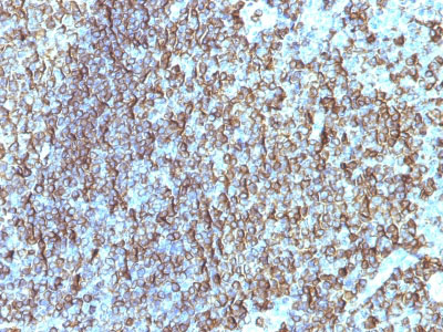
Application 
| WB, IHC, IF, FC |
|---|---|
| Primary Accession | P01911 |
| Other Accession | 3123, 534322 |
| Reactivity | Human, Mouse, Monkey |
| Host | Mouse |
| Clonality | Monoclonal |
| Isotype | Mouse / IgG2b, kappa |
| Clone Names | SPM288 |
| Calculated MW | 28kDa (beta chain) |
| Gene ID | 3123 |
|---|---|
| Other Names | HLA class II histocompatibility antigen, DRB1-15 beta chain, DW2.2/DR2.2, MHC class II antigen DRB1*15, HLA-DRB1, HLA-DRB2 |
| Storage | Store at 2 to 8°C.Antibody is stable for 24 months. |
| Precautions | HLA-DRB (MHC II) Antibody - With BSA and Azide is for research use only and not for use in diagnostic or therapeutic procedures. |
| Name | HLA-DRB1 (HGNC:4948) |
|---|---|
| Function | A beta chain of antigen-presenting major histocompatibility complex class II (MHCII) molecule. In complex with the alpha chain HLA- DRA, displays antigenic peptides on professional antigen presenting cells (APCs) for recognition by alpha-beta T cell receptor (TCR) on HLA-DRB1-restricted CD4-positive T cells. This guides antigen-specific T-helper effector functions, both antibody-mediated immune response and macrophage activation, to ultimately eliminate the infectious agents and transformed cells (PubMed:15265931, PubMed:16148104, PubMed:22327072, PubMed:27591323, PubMed:29884618, PubMed:31495665, PubMed:8642306). Typically presents extracellular peptide antigens of 10 to 30 amino acids that arise from proteolysis of endocytosed antigens in lysosomes (PubMed:8145819). In the tumor microenvironment, presents antigenic peptides that are primarily generated in tumor- resident APCs likely via phagocytosis of apoptotic tumor cells or macropinocytosis of secreted tumor proteins (PubMed:31495665). Presents peptides derived from intracellular proteins that are trapped in autolysosomes after macroautophagy, a mechanism especially relevant for T cell selection in the thymus and central immune tolerance (PubMed:17182262, PubMed:23783831). The selection of the immunodominant epitopes follows two processing modes: 'bind first, cut/trim later' for pathogen-derived antigenic peptides and 'cut first, bind later' for autoantigens/self-peptides (PubMed:25413013). The anchor residue at position 1 of the peptide N-terminus, usually a large hydrophobic residue, is essential for high affinity interaction with MHCII molecules (PubMed:8145819). |
| Cellular Location | Cell membrane; Single-pass type I membrane protein. Endoplasmic reticulum membrane; Single-pass type I membrane protein. Lysosome membrane; Single-pass type I membrane protein. Late endosome membrane; Single-pass type I membrane protein. Autolysosome membrane Note=The MHC class II complex transits through a number of intracellular compartments in the endocytic pathway until it reaches the cell membrane for antigen presentation (PubMed:18305173). Component of immunological synapses at the interface between T cell and APC (PubMed:29884618). |
| Tissue Location | Expressed in professional APCs: monocyte/macrophages, dendritic cells and B cells (at protein level) (PubMed:19830726, PubMed:23783831, PubMed:31495665). Expressed in thymic epithelial cells (at protein level) (PubMed:23783831) |