General Info
| Host: | Mouse |
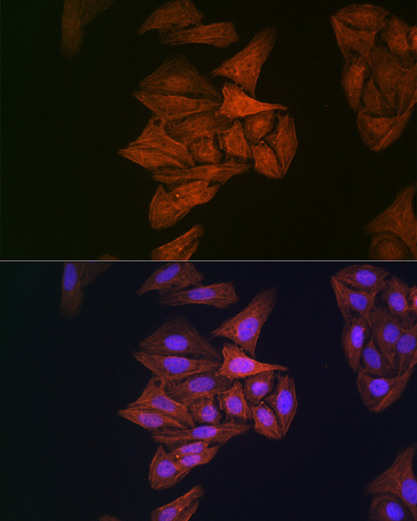
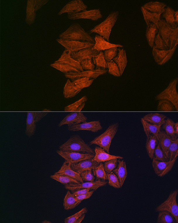
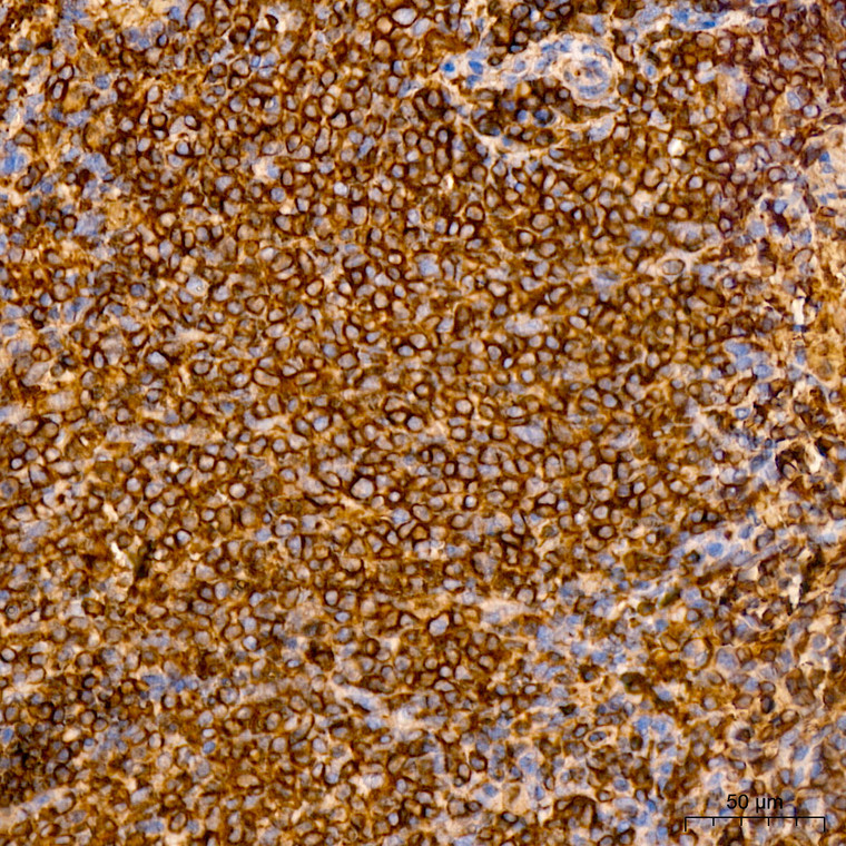
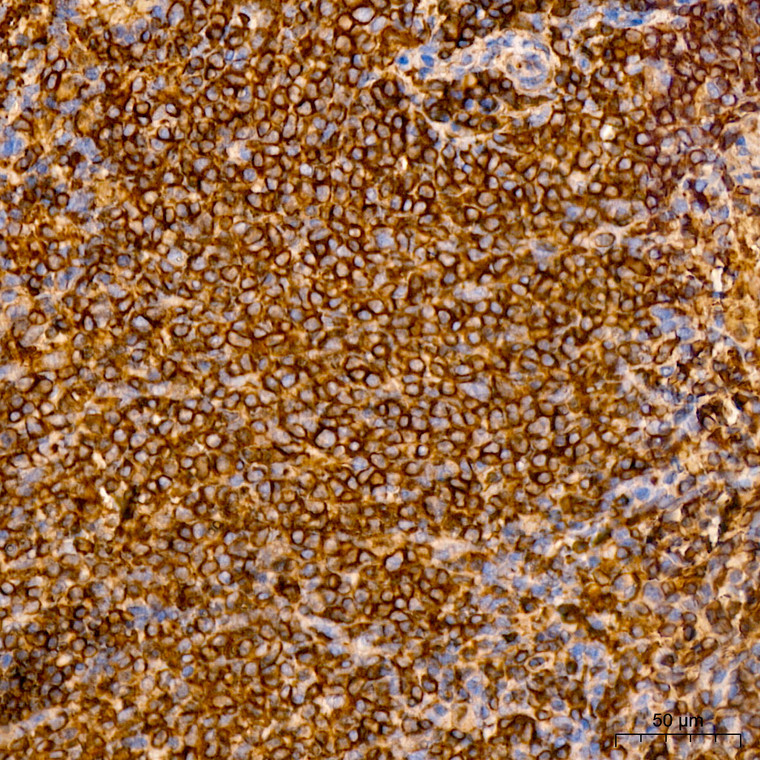
| Applications: | WB/IHC/IF |
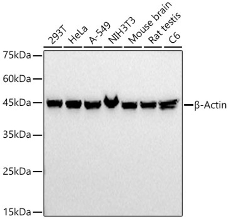
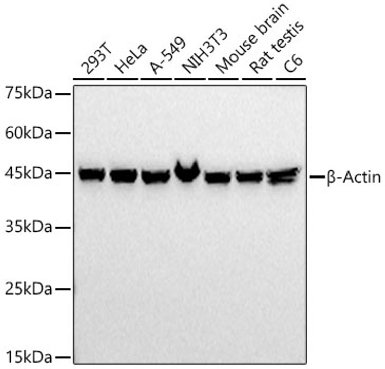
| Reactivity: | Human/Mouse/Rat |
| Note: | STRICTLY FOR FURTHER SCIENTIFIC RESEARCH USE ONLY (RUO). MUST NOT TO BE USED IN DIAGNOSTIC OR THERAPEUTIC APPLICATIONS. |
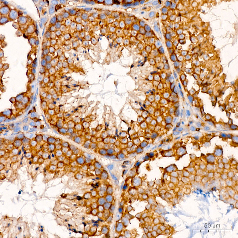
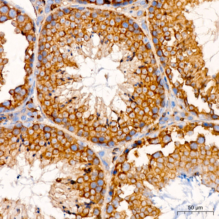
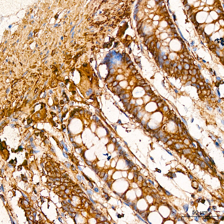
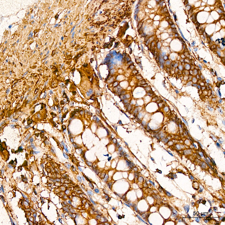
| Short Description: | Mouse monoclonal antibody anti-Beta-Actin is suitable for use in Western Blot, Immunohistochemistry and Immunofluorescence research applications. |
| Clonality: | Monoclonal |
| Clone ID: | S3MR |
| Conjugation: | Unconjugated |
| Isotype: | IgG1k |
| Formulation: | PBS with 0.01% Thimerosal, 50% Glycerol, pH7.3. |
| Purification: | Affinity purification |
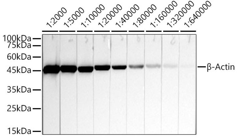
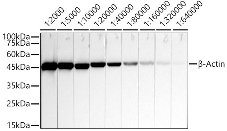
| Dilution Range: | WB 1:2000-1:20000IHC-P 1:50-1:200IF/ICC 1:50-1:200 |
| Storage Instruction: | Store at-20°C for up to 1 year from the date of receipt, and avoid repeat freeze-thaw cycles. |
Information
| Gene Symbol: | ACTB |
| Gene ID: | 60 |
| Uniprot ID: | ACTB_HUMAN |
| Immunogen: | Fusion protein of human Beta-Actin |
| Immunogen Sequence: | Email for sequence |
Description
| Post Translational Modifications | ISGylated. Oxidation of Met-44 and Met-47 by MICALs (MICAL1, MICAL2 or MICAL3) to form methionine sulfoxide promotes actin filament depolymerization. MICAL1 and MICAL2 produce the (R)-S-oxide form. The (R)-S-oxide form is reverted by MSRB1 and MSRB2, which promote actin repolymerization. Monomethylation at Lys-84 (K84me1) regulates actin-myosin interaction and actomyosin-dependent processes. Demethylation by ALKBH4 is required for maintaining actomyosin dynamics supporting normal cleavage furrow ingression during cytokinesis and cell migration. Methylated at His-73 by SETD3. Methylation at His-73 is required for smooth muscle contraction of the laboring uterus during delivery. Actin, cytoplasmic 1: N-terminal cleavage of acetylated methionine of immature cytoplasmic actin by ACTMAP. Actin, cytoplasmic 1, N-terminally processed: N-terminal acetylation by NAA80 affects actin filament depolymerization and elongation, including elongation driven by formins. In contrast, filament nucleation by the Arp2/3 complex is not affected. (Microbial infection) Monomeric actin is cross-linked by V.cholerae toxins RtxA and VgrG1 in case of infection: bacterial toxins mediate the cross-link between Lys-50 of one monomer and Glu-270 of another actin monomer, resulting in formation of highly toxic actin oligomers that cause cell rounding. The toxin can be highly efficient at very low concentrations by acting on formin homology family proteins: toxic actin oligomers bind with high affinity to formins and adversely affect both nucleation and elongation abilities of formins, causing their potent inhibition in both profilin-dependent and independent manners. |
| Function | Actin is a highly conserved protein that polymerizes to produce filaments that form cross-linked networks in the cytoplasm of cells. Actin exists in both monomeric (G-actin) and polymeric (F-actin) forms, both forms playing key functions, such as cell motility and contraction. In addition to their role in the cytoplasmic cytoskeleton, G- and F-actin also localize in the nucleus, and regulate gene transcription and motility and repair of damaged DNA. Part of the ACTR1A/ACTB filament around which the dynactin complex is built. The dynactin multiprotein complex activates the molecular motor dynein for ultra-processive transport along microtubules. |
| Protein Name | Actin - Cytoplasmic 1Beta-Actin Cleaved Into - Actin - Cytoplasmic 1 - N-Terminally Processed |
| Database Links | Reactome: R-HSA-1445148Reactome: R-HSA-190873Reactome: R-HSA-196025Reactome: R-HSA-2029482Reactome: R-HSA-3214847Reactome: R-HSA-389957Reactome: R-HSA-390450Reactome: R-HSA-3928662Reactome: R-HSA-3928665Reactome: R-HSA-418990Reactome: R-HSA-437239Reactome: R-HSA-4420097Reactome: R-HSA-445095Reactome: R-HSA-446353Reactome: R-HSA-5250924Reactome: R-HSA-5626467Reactome: R-HSA-5663213Reactome: R-HSA-5663220Reactome: R-HSA-5674135Reactome: R-HSA-5689603Reactome: R-HSA-5696394Reactome: R-HSA-6802946Reactome: R-HSA-6802948Reactome: R-HSA-6802952Reactome: R-HSA-6802955Reactome: R-HSA-8856828Reactome: R-HSA-9035034Reactome: R-HSA-9649948Reactome: R-HSA-9656223Reactome: R-HSA-9662360Reactome: R-HSA-9662361Reactome: R-HSA-9664422Reactome: R-HSA-983231 |
| Cellular Localisation | CytoplasmCytoskeletonNucleusLocalized In Cytoplasmic Mrnp Granules Containing Untranslated Mrnas |
| Alternative Antibody Names | Anti-Actin - Cytoplasmic 1 antibodyAnti-Beta-Actin Cleaved Into - Actin - Cytoplasmic 1 - N-Terminally Processed antibodyAnti-ACTB antibody |
Information sourced from Uniprot.org
12 months for antibodies. 6 months for ELISA Kits. Please see website T&Cs for further guidance


